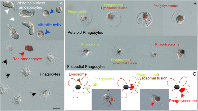
Figure 2. Neutral Red for live cell imaging of the phagocytic activity of exposed immune cells.
(A) The pH-indicator dye NR is concentrated in lysosomes forming typical small red vesicles within all three different cell types of freely circulating immune cells present in Paracentrotus lividus (phagocytes, amoebocytes and vibratile cells), most evident in the vibratile cells. Cell types are indicated by captions of different colors and corresponding pointing arrows as described in Fig. 1. (B) Petaloid and filopodial phagocytes show areas of high lysosomal and phagocytic activity in which NR became concentrated. (C) Schematic model and demonstrative pictures representing an immune cell undergoing phagosomal maturation. Arrows indicate lysosomes (orange arrows), phagosomal/lysosomal fusion (yellow arrows), phagolysosome (red arrows). Bar 5 μm.

