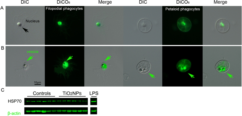
Figure 3. Dihexyloxacarbocyanine iodide to monitor lysosomal internal membrane stability of TiO2 exposed immune cells.

(A) The green fluorescent membrane dye DiOC6 is detected within the cytoplasm, as a dense signal around the nuclei of both the non-activate filopodial and petaloid phagocytes. (B) A few cells showed a growing network of vesicles (green arrows), validating a phagocytic activity in progress. (C) Representative image of the HSP70 levels of 6 control specimens, 6 specimens exposed to TiO2NP and 1 to LPS, evaluated by immunoblotting. Contrary to LPS exposure, TiO2NP exposure do not stimulate the activation of the HSP70-dependent stress response in the sea urchin immune cells.
