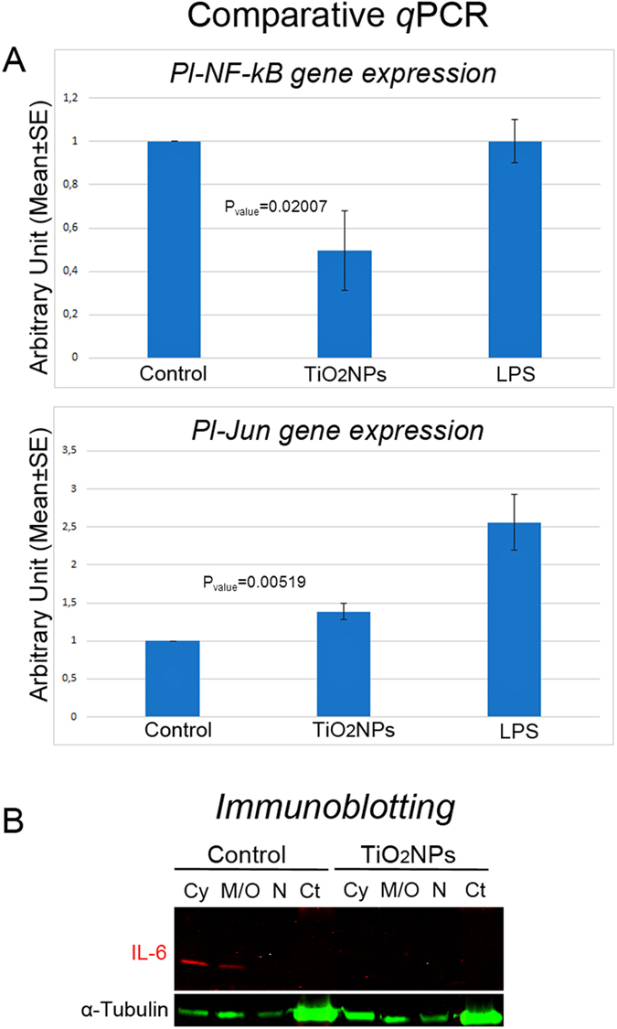
Figure 6. Signal transduction downstream to p38 MAPK in response to TiO2 nanoparticles.

(A) Comparative qPCR analysis show levels of expression of Pl-NF-kB gene 2-fold lower than those measured in controls (injected only with ASW) (upper panel), while the transcript of Pl-Jun showed a weak increase in its levels (lower panel). Levels are expressed in arbitrary units as fold increase compared to controls assumed as 1, using the endogenous gene Pl-Z12-1 for normalization. Results were compared to those obtained in response to LPS (2 μg/ml in ASW, 24 h): in this case, the levels of expression of Pl-NF-kB gene were found comparable to controls while the transcript of Pl-Jun was found strongly over-expressed (2.56 ± 0.71-fold). Each bar represents the mean of three independent experiments ±SE. (B) Immunoblotting analysis with anti-IL6 subcellular fractions of sea urchin immune cells. Cy: cytosol; M/O: membrane/organelles; N: nuclei; Ct: cytoskeleton.
