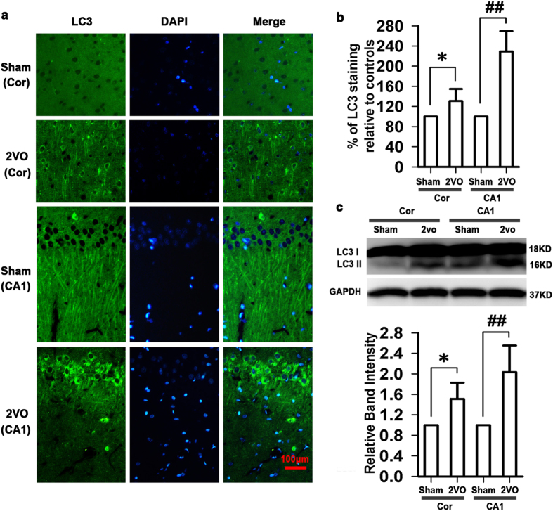
Figure 2. Activation of autophagy in cortex and hippocampal CA1 area under chronic cerebral hypoperfusion.
(a) Representative photomicrographs of immunohistochemical staining with anti-LC3 antibody in cortex (scale bar, 100 μm). Five weeks after induction of hypoperfusion, the LC3 immunoreactivity was slightly but significantly increased in cortex, however, a robust increase in the LC3 immunoreactivity was observed in hippocampal CA1 area (n = 4 in each group). (b) Quantitative analysis of the LC3 immunoreactivity. (c) The protein expression of LC3 II in cortex and hippocampal CA1 area (n = 4 in each group). Blots shown have been cropped to fit space requirements and run under the same experimental conditions. *P < 0.05 vs sham-operated rats (cortex), ##P < 0.01 vs sham-operated rats (hippocampal CA1 area).

