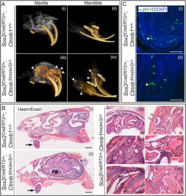
Figure 1. Postnatal expression of activated β-catenin in SOX2+ dental stem cells results in abnormal structures resembling odontoma.
(A) Three-dimensional reconstruction of microCT scans of the maxilla and mandible of control Sox2CreERT2/+; Ctnnb1+/+ and mutant Sox2CreERT2/+; Ctnnb1lox(ex3)/+ animals at 5 months of age, 3 months after tamoxifen-induced CreERT2 activation. Mutant animals display multiple ectopic odontogenic structures of similar density to enamel and dentine, concentrated around the base of the incisors (arrowheads). (B) Haematoxylin and eosin-stained sagittal sections through a control decalcified head at 5 months of age (i), compared to a Sox2CreERT2/+; Ctnnb1lox(ex3)/+ mutant (ii–viii), 3 months following tamoxifen induction. Incisors are indicated by arrows and asterisk in (ii). Note the large malformation in the mutant outlined by dashed line in (ii). The asterisk denotes part of the normal incisor. Higher magnification images of malformations are shown in iii-viii. Arrowheads indicate the boundaries of malformation circumscription (iii), cortical thinning (iv), duct-like foci (v) and a tooth-like structure with multiple cusps (vi). Bracket in (vii) indicates normal orientation of epithelial and mesenchymal layers within a tooth-like structure (amb, ameloblasts; em, enamel matrix; de, dentine; pde, predentine; ob, odontoblasts). Irregular mineralization, osteodentine and dysplastic dentine, consistent with complex odontoma is shown in (viii), arrowheads. (C) Immunofluorescence staining with antibodies against phospho-histone-H3 to mark proliferating cells showing the normal proliferation pattern in the cervical loop of a normal incisor (i) and a similar pattern in an individual tooth-like structure from within the malformation (ii). Scale bars in B(i) = 1000 μm, B(ii–vii) and C = 100 μm.

