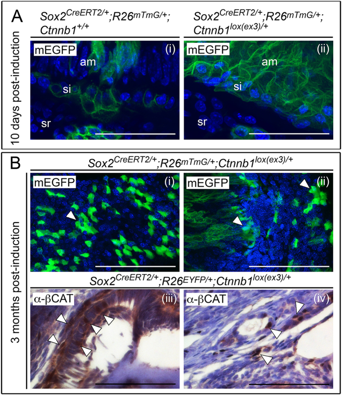
Figure 2. Genetic tracing of postnatal SOX2+ dental stem cells shows that structures resembling odontoma are heterogeneous in origin.
(A) Lineage-tracing of SOX2+ cells at the base of the incisor by membrane-EYFP fluorescence in postnatal Sox2CreERT2/+; R26mTmG/+; Ctnnb1+/+ (i) andlox(ex3)/+ (ii) animals, 10 days following tamoxifen induction (sr, stellate reticulum; si, stratum intermedium; am, ameloblasts). Nuclei are counterstained with DAPI (blue). (B) Lineage tracing by fluorescence of membrane-EGFP positive cells in mature structures from malformations in Sox2CreERT2/+; R26mTmG/+; Ctnnb1lox(ex3)/+ mice, reveals a proportion of EGFP positive cells (green, arrowheads to positive clusters of cells, i-ii) as well as EGFP negative cells. Nuclei are counterstained with DAPI (blue). Staining with anti-β-catenin antibodies (α-βCAT) showing individual cells with nucleo-cytoplasmic accumulation of β-catenin (arrowheads, iii-iv), consistent with cells sustaining the β-catenin mutation. Sections are counterstained with haematoxylin. Scale bars = 50 μm in A, 100 μm in (B).

