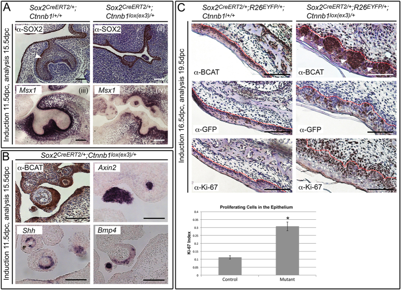
Figure 3. Embryonic induction of constitutive active β-catenin in SOX2+ embryonic progenitors reveals a non-cell autonomous mechanism of proliferation.
(A) Tamoxifen induction at 11.5 dpc and analysis at 15.5 dpc reveals formation of multiple tooth buds in Sox2CreERT2/+; Ctnnb1lox(ex3)/+ embryos. Immunohistochemistry with SOX2 antibodies marks the epithelium in Sox2CreERT2/+; Ctnnb1+/+ control (i) and Sox2CreERT2/+; Ctnnb1lox(ex3)/+ mutant (ii). Note the uniform SOX2 distribution in the mutant, not exhibiting the stronger lingual expression observed in the control (arrowhead). Sections counterstained with haematoxylin. In situ hybridisation (ISH) with riboprobes against the BMP target Msx1, marks condensing odontogenic mesenchyme in control (iii), and mutant (iv) embryos, confirming Msx1 expression surrounding the abnormal buds in the mutant. (B) Sox2CreERT2/+; Ctnnb1lox(ex3)/+ embryos show strong nucleo-cytoplasmic accumulation of β-catenin in foci, and aberrant expression of Shh and Bmp4 as seen by ISH. Ectopic activation of the WNT pathway is confirmed through expression of Axin2. (C) Tamoxifen induction at 16.5 dpc and analysis at 19.5 dpc results in discrete foci accumulating β-catenin in the OE at the level of the second molar, in Sox2CreERT2/+; R26EYFP/+; Ctnnb1lox(ex3)/+ mice (arrowheads). Immunohistochemistry with anti-GFP antibodies (α-GFP) tracing the fate of CreERT2-expressing cells, revealing GFP positive cells within β-catenin accumulating regions (consecutive sections). Immunohistochemistry with antibodies against Ki-67 marks proliferating cells. Dotted red lines mark epithelial borders. Graph of epithelial mitotic index quantification in control (Sox2CreERT2/+; R26EYFP/+; Ctnnb1+/+) and mutant (Sox2CreERT2/+; R26EYFP/+; Ctnnb1lox(ex3)/+) embryos at 19.5 dpc. Error bars represent the standard deviation. Ki-67 increase is significant in mutants (unpaired t-test, P < 0.0001). Scale bars = 100 μm.

