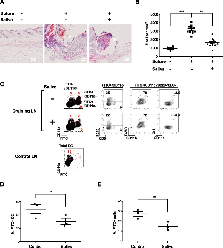
Figure 1.

Dual effect of tick saliva in vivo on the recruitment of leukocyte infiltrates to skin and dendritic cells to draining lymph nodes. A surgical monofilament suture was positioned on the back of a C57BL/6 mouse and tick saliva or a control solution was intradermally injected once a day for 3 days into the suture area. The skin at the sutured area was then stained with FITC solution and mice were killed 24 h later to analyze the skin and lymph nodes. Data were obtained from two independent experiments including three mice per condition. (A) Representative hematoxylin and eosin staining (H&E) of histological sections of skin biopsies. B Density of infiltrated leukocytes in skin biopsies was estimated by counting the total number of nuclei within histological samples of skin sections, of similar size and including the sutured-damaged area, using Image J software (two mice per condition, 10 sections per mouse). Statistically significant differences between groups were assessed with the ANOVA (Kruskal-Wallis) test and Dunn’s multiple comparison post test, * P < 0.05 and ** P < 0.005. C Contour plots of resident DCs (FITC−CD11c+), migrating DCs (FITC+ CD11C+) and other migrating cells (FITC+ CD11C−) in the draining lymph nodes from a control skin area (no FITC, Control LN) and from a sutured areas injected with saliva or control solution (Draining LN). FITC+ CD11C− cells include migrating B cells (B220+), macrophages (CD11b+F4/80+) and granulocytes (CD11b+Gr1+). D-E The relative proportion of total migratory cells (FITC+) and migratory DCs (FITC+ CD11C+) in lymph nodes draining the suture area was determined in the absence or presence of saliva. The mean percentage of migratory cells in each condition is indicated with a line and each point represents one mouse. Statistically significant differences between the two groups were determined by the Student’s t test, *P < 0.05 and **P < 0.005.
