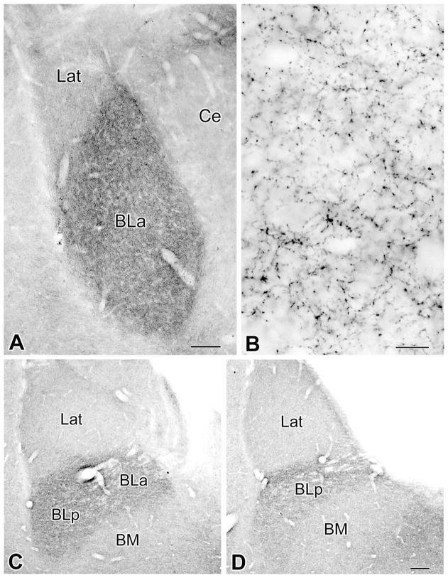
Figure 1.
Innervation of the amygdala by cholinergic VAChT+ axons. A: Low-power light micrograph of VAChT immunoreactivity at the Bregma –2.3 level (Paxinos and Watson, 1997). Note that the density of VAChT+ axons in the BLa is much greater than in the lateral nucleus (Lat) and that the lateral subdivision of the central nucleus (Ce) contains very few VAChT+ axons. B: Higher power light micrograph of VAChT+ axons in the BLa at the level of A. C,D: Low-power light micrographs of VAChT immunoreactivity at the Bregma –3.3 level (C) and Bregma –4.0 level (D). Note that the density of VAChT+ axons in the BLa and BLp is much greater than in the lateral (Lat) or basomedial nucleus (BM). Scale bars = 150 μm in A; 10 μm in B; 150 μm in D (applies to C,D).

