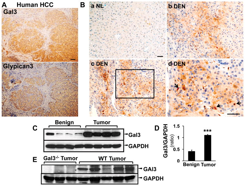
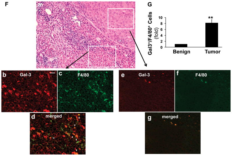
Figure 3. Galectin 3 is upregulated in HCC tumor cells and tumor associated macrophages.
Sequential sections from human HCC patients were used for immunohistochemistry staining targeting galectin 3 and glypican 3 (A). The image showed that the galectin 3 positive areas were highly identical with the glypican 3 positive areas in human HCC. The immunohistochemistry staining for galectin 3 was then conducted in the tissues from normal (NL) and wt DEN treated mice (B). Galectin 3 was highly expressed in DEN induced tumors (b–d), mainly in hepatoma cells. The high magnification image (B, d) depicted tumor-associated macrophages also expressing galectin 3 (arrow). Normal hepatocytes in control liver did not express galectin 3 (B, a). Western blot analysis and ImageJ quantification showed that galectin 3 expression was significantly higher in the tumor tissues (C, D. N=4, ***p<0.001). Western blot assay was also conducted on the wt and galectin 3−/− tumors for antibody validation (E). No bands were detected in ~30kDa area in the knockout tumors and strong signals were shown in the wt tumors. Immunofluorescence and confocal microscopy on consecutive sections to the H&E-stained sections was performed to assess galectin 3 (red) and F4/80 (marker for tumor associated macrophages TAM, green, F). Analyzing the areas of HCC bordering the surrounding parenchyma, clusters of TAM co-expressed galectin 3 and F4/80 (b–d, arrowhead, Bar=50 μm) whereas no expression was seen in the normal hepatocytes and F4/80 positive cells were rare in the normal tissue (e–g). The tumor tissue carried a significant higher number of galectin 3+/F4/80+ cells (G, **p<0.01)


