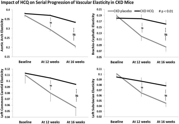
Fig 6. Quantitative analysis of vascular elasticity in CKD groups.
Quantification of vascular wall elasticity as visualized by M-mode intravital ultrasound echography using VisualSonics Vevo 770 in abdominal aortas. The elasticity of the major vessels, i.e., the movement of vessel wall between the systole and diastole, as well as blood velocity within these vessels were significantly better in CKD mice treated with HCQ. *P < 0.05 compared with CKD placebo mice. AA, aortic arch; BCA, brachio-cephalic artery; LCCA, left common carotid artery; LSA, left subclavian artery.

