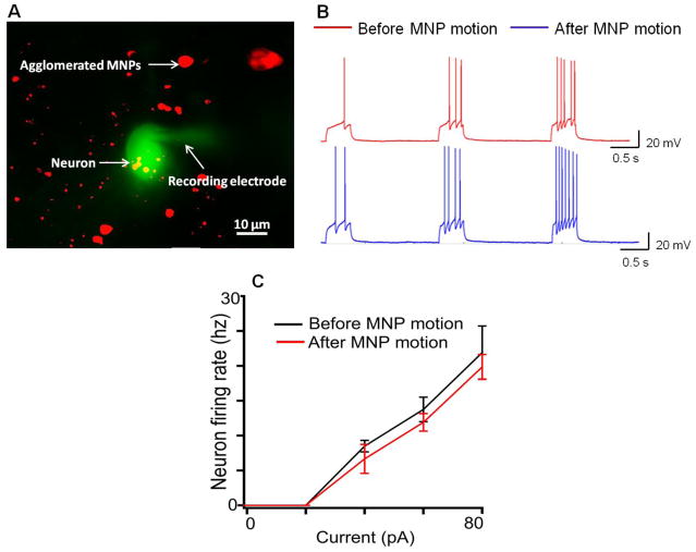
Figure 2.
Functional health of brain tissue after MNP motion. (A) Recording from a mitral cell in the olfactory bulb after the slices treated with MNPs were subjected to a magnetic field. The recording electrode contained the fluorescent dye Alexa-488 (green), which diffuses into the neuron during the recording. The MNPs contained a fluorophore Texas-Red (red). Note this is a total summed two wavelength images (B). Current-clamp recordings in mitral cells before (red) and after magnet induced MNP movement (blue). Increasing depolarizing current pulses (not shown) elicited action potentials in both control and treated neurons. (C) In the range of depolarizing current used, the frequency of neuronal firing increased linearly and it was comparable for different constant current stimuli before (black) and after MNP motion (red).

