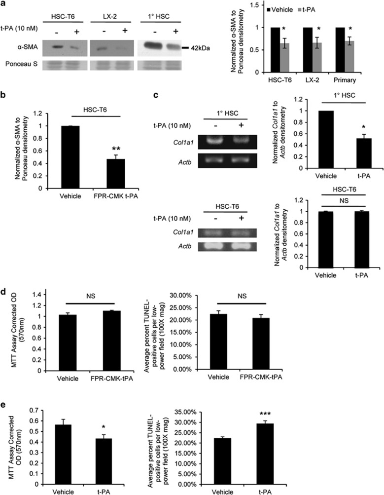
Figure 1.
t-PA inhibits HSC activation in cultured cells. (a) Representative α-SMA western blots on HSC-T6, LX-2, or primary rat HSCs treated with exogenous t-PA for 24 h, using pooled whole-cell lysates (n=2–3/well); quantification on right (n=5 independent experiments per cell source). (b) Quantifications of western blots for α-SMA on lysates from HSC-T6 cells treated for 24 h with protease-inactivated t-PA (n=4). (c) RT-PCR for Col1a1 mRNA expression in primary HSCs or HSC-T6 cells after 24 h t-PA treatment; quantification on right (n=3 and 4, respectively). (d and e) HSC-T6 cells stimulated with protease-inactivated t-PA (d) or t-PA (e) for 48 h before measuring mitochondrial function (MTT reduction assay; n=3) or apoptosis (TUNEL labeling). Error bars±s.e.m. *P<0.05, **P<0.01, ***P<0.001, Student's paired (a and b) or unpaired (c–e) t-test.

