Figure 3.
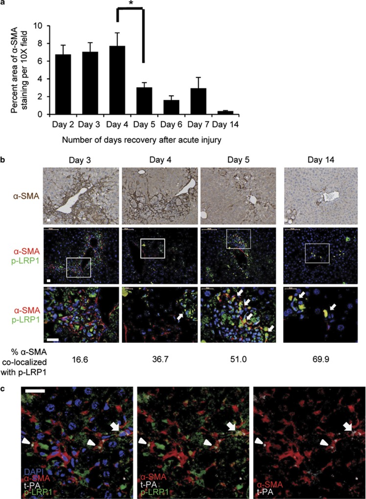
LRP1 is tyrosine phosphorylated in HSCs during resolution of acute hepatic injury in vivo. (a) Quantification of α-SMA immunohistochemistry on tissue sections from WT mice after acute CCl4 injury as described in MATERIALS AND METHODS (× 10 images, minimum 10). *P<0.05, one-way ANOVA with post-test comparisons; only sequential differences denoted. Error bars±s.e.m. (b) Standard immunohistochemistry (upper panels, α-SMA) or double Qdot-labeled immunohistochemistry for p-LRP1 and α-SMA (middle and lower panels), with percent α-SMA/p-LRP co-localization area of total α-SMA staining indicated below each time point. Arrows indicate co-localization. Bottom panels are higher magnifications of boxed regions in the middle panels. Days indicated are post-CCL4. (c) Qdot-labeled immunohistochemistry for α-SMA, t-PA, and p-LRP1 on day 4 post-CCL4. Selected channels separated from the same image shown for comparison. Arrow indicates triple co-location, arrowhead indicates α-SMA/t-PA co-localization. Scale bars, 20 μm.

