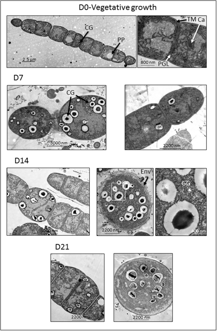
FIGURE 1.
Electron micrographs of ultra-thin sections of filaments and akinetes of Aphanizomenon ovalisporum captured at different time points (D0, day 0; D7, day 7; D14, day 14; and D21, day 21) after akinete-induction (transfer to K+-depleted medium). Ca, carboxysome; CG, cyanophycin globule; GG, glycogen globule; Env, envelope; PP, polyphosphate body; TM, thylakoid membrane. The right electron micrograph of D14 lane is the magnified part indicated by a white frame of the middle micrograph. A scale bar is presented for each micrograph.

