
Figure 1. Cell viability and apoptosis of ARPE-19 cells cultured with Angiotensin II as measured with CCK-8 and Annexin-V FITC/PI assay.
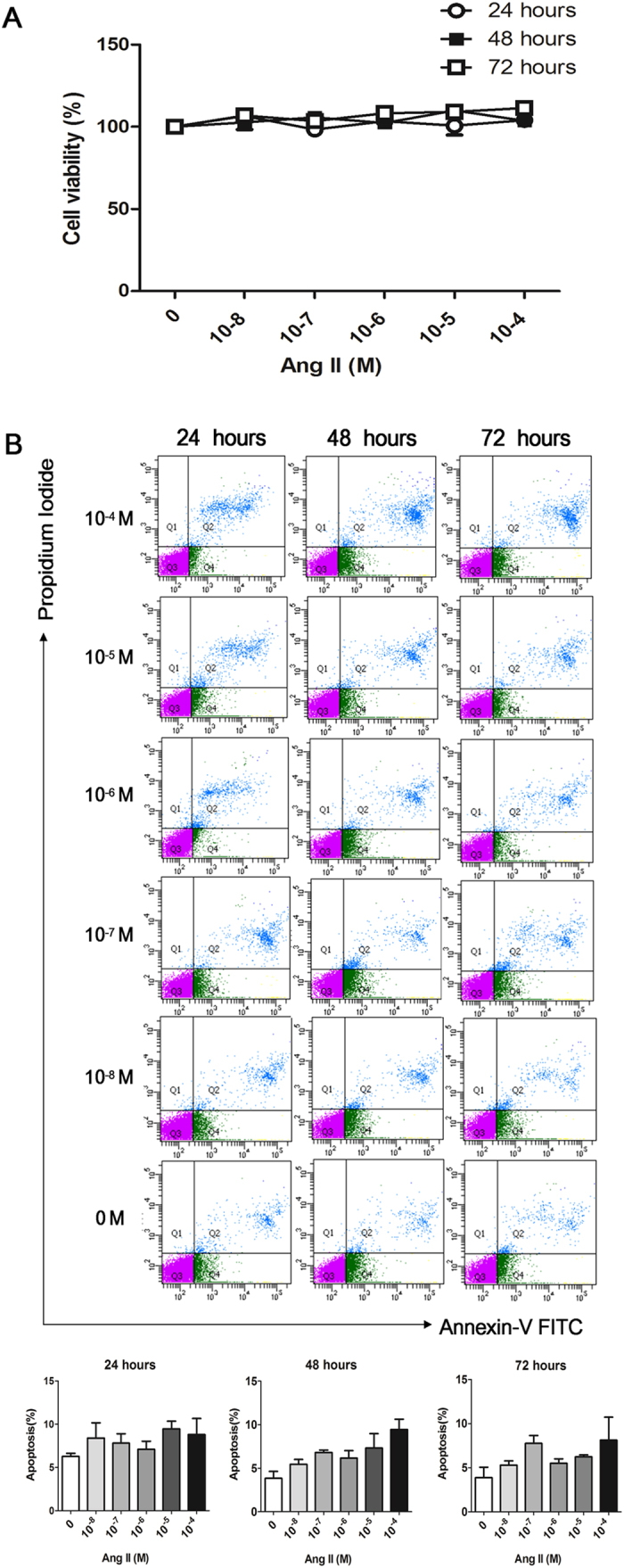
(A) ARPE-19 cells were cultured with various concentrations (10−8, 10−7, 10−6, 10−5 and 10−4 M) of Ang II for 24, 48 and 72 hours. Incubation with Ang II did not affect the viability of ARPE-19 cells compared with cells cultured without Ang II. Viability of each treatment group was measured compared to cells cultured without Ang II (0 M). There was no significant difference in cellular viability among the treatment groups at any time point. The data were normalized to the control group (0 M) (p > 0.05, n = 3). (B) The lower left quadrant (Q3) represents viable cells (Annexin-FITC and PI negative). The upper right quadrant (Q2) represents late apoptotic cells (Annexin-FITC and PI positive). There was no significant difference of cell apoptosis among the cells cultured with various concentrations of Ang II for 24, 48 and 72 hours. The data were shown as mean ± SEM (p > 0.05, n = 4).