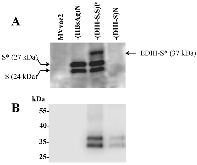
Figure 3.
Analysis of expression of the hybrid DIII-HBsAg in lysates from recombinant MV-infected cells (indicated on the top of each panel). In (A) SDS-PAGE under non-reducing conditions using a monoclonal antibody against HBsAg (the S antigen); the molecular weight of the two forms of S are shown to the left and the molecular weight of the hybrid DIII-S to the right. The asterisk indicates glycosylated forms. In (B) SDS-PAGE under reducing conditions using a monoclonal antibody against dengue envelope protein domain III.

