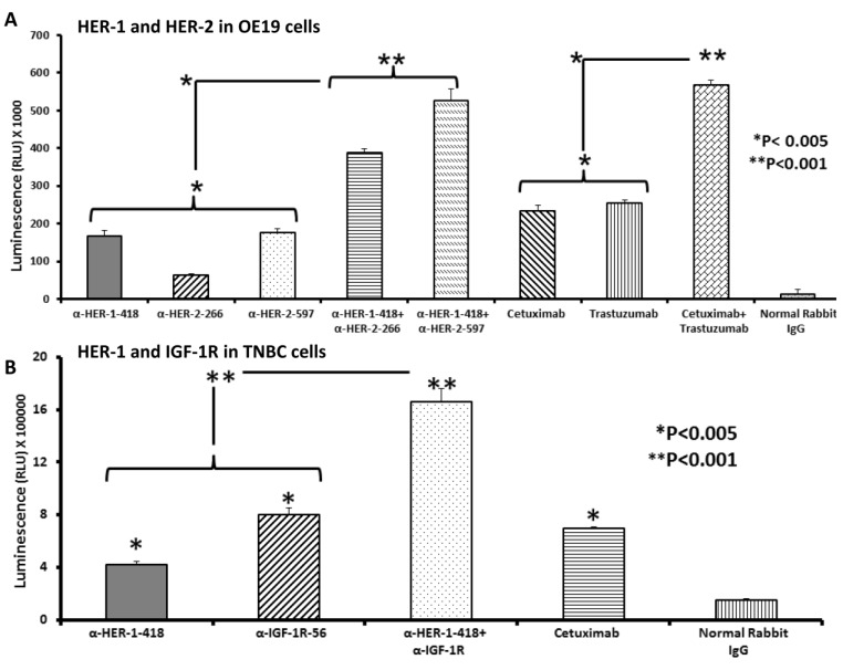
Figure 4.
Effects of co-targeting HER-1 and HER-2 or HER-1 and IGF-1R on induction of apoptosis in EC or TNBC. OE19 or MDA-MB-231 cancer cells were plated in 96-well plates and treated with peptide vaccine antibodies for 24 h prior to cell lysis. Apoptosis (directly proportional to amount of luminescence produced) was measured using the Caspase Glo 3/7 kit (Promega, Madison, WI, USA). After 24 h of treatment, caspase glo reagent was added, and plates were incubated for 3 h before being read on a luminometer. Normal rabbit IgG was used as a negative control; cetuximab and trastuzumab were positive controls. Results are representative of two independent experiments performed in triplicate. Error bars represent SD of the mean. In EC cells (A), single peptide vaccine antibody treatment significantly increased apoptosis over negative control (* p < 0.005). The combinations of anti-HER-1-418 and HER-2-266 or anti-HER-1-418 and HER-2-597 showed significantly higher levels of apoptosis than single treatment alone (* p < 0.005). TNBC cells (B) showed single peptide vaccine antibody treatment significantly increased apoptosis over negative control (* p < 0.005), and the combination of anti-HER-1-418 and anti-IGF-1R-56 showed significantly higher levels of apoptosis compared to single treatment alone (** p < 0.001).

