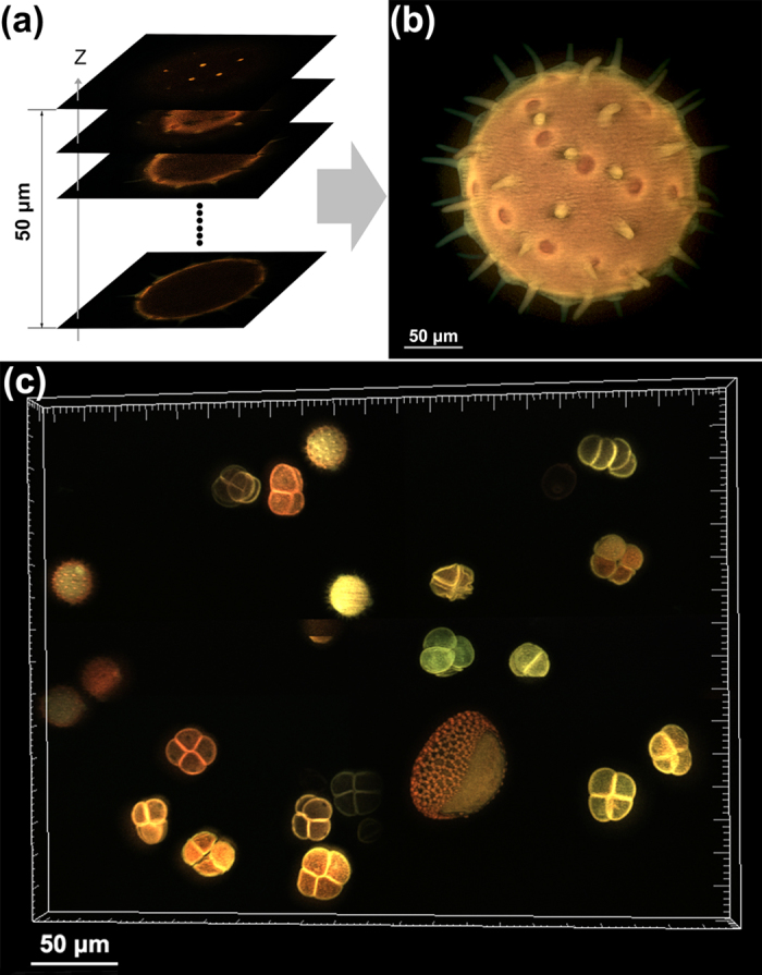
Figure 2. Optical sectioning images of the thick volume structure of different pollen grains.

The auto-fluorescence signals of the pollen grains are excited by 405 nm light and collected by 20 × objective. (a) A sequence of optically sectioned images. (b) The maximum-intensity projection of all the planes along Z-axis. (c) The maximum-intensity projection of differently shaped and colorful pollen grains (Supplementary Movie 1).
