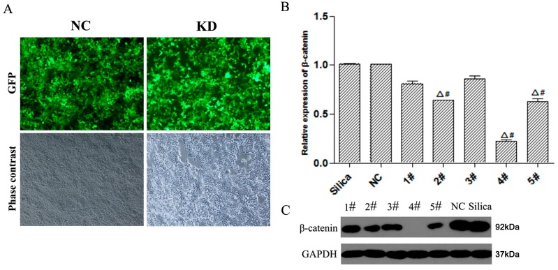
Figure 1.
Infection of MLE-12 cells with non-silenced control (NC) or Lv-shβ-catenin (KD). (A) Cells were infected with NC or Lv-shβ-catenin. Phase contrast image and GFP expression under a fluorescent microscope was taken after 72 h. (B) After MLE-12 cells were treated with Lv-shβ-catenin, mRNA level of β-catenin was detected by realtime-PCR by using -ΔΔ Ct method. Data are mean ± SEM (n = 3). (C) Protein level of β-catenin was detected by western blot. Cells of each group were incubated with silica. Lanes 1–5: Lv-shβ-catenin-1, -2, -3, -4, -5, respectively. Δ p < 0.05, compared to the silica group; # p < 0.05, compared to the NC group.

