Figure 4.
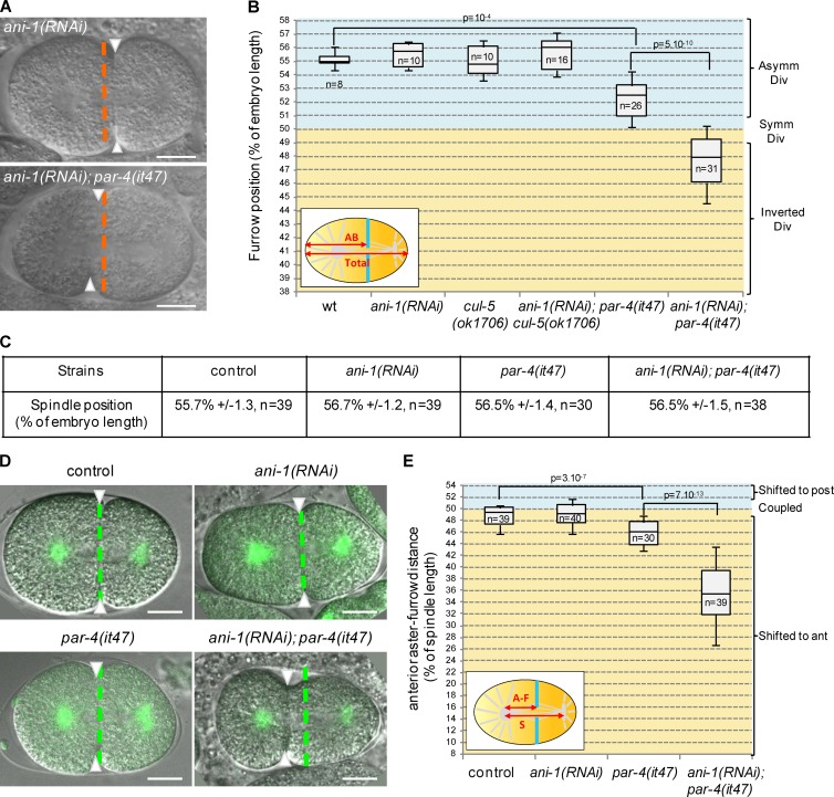
Loss of PAR-4 and ANI-1 leads to strong furrow mispositioning. (A and B) ani-1(RNAi); par-4(it47) embryos show strong furrow mispositioning. DIC images (A) and quantification of furrow position (B) in dividing one-cell embryos of the indicated genotypes. Furrow position was measured when the furrow reached its most anterior position. Orange dashed lines correspond to the embryo center and arrowheads to furrow position. See also Fig. S2 (A and D) and Video 3. (C) Position of the spindle center at the onset of cytokinesis in one-cell embryos of the indicated genotypes (all strains also express an α-tub::YFP transgene). (D and E) Furrow and spindle positions are strongly uncoupled in ani-1(RNAi); par-4(it47) embryos. Superposed confocal sections and DIC images (D) and quantification of furrow/spindle coupling (E) in dividing one-cell embryos of the indicated genotypes (all strains also express an α-tub::YFP transgene [green]). Furrow position was measured when the furrow reached its most anterior position. Green dashed lines correspond to the spindle center and arrowheads to furrow position. Experiment was performed at 20°C. See also Fig. S2 (B and C) and Video 4. Bars, 10 µm. P-values from Student’s t test.

