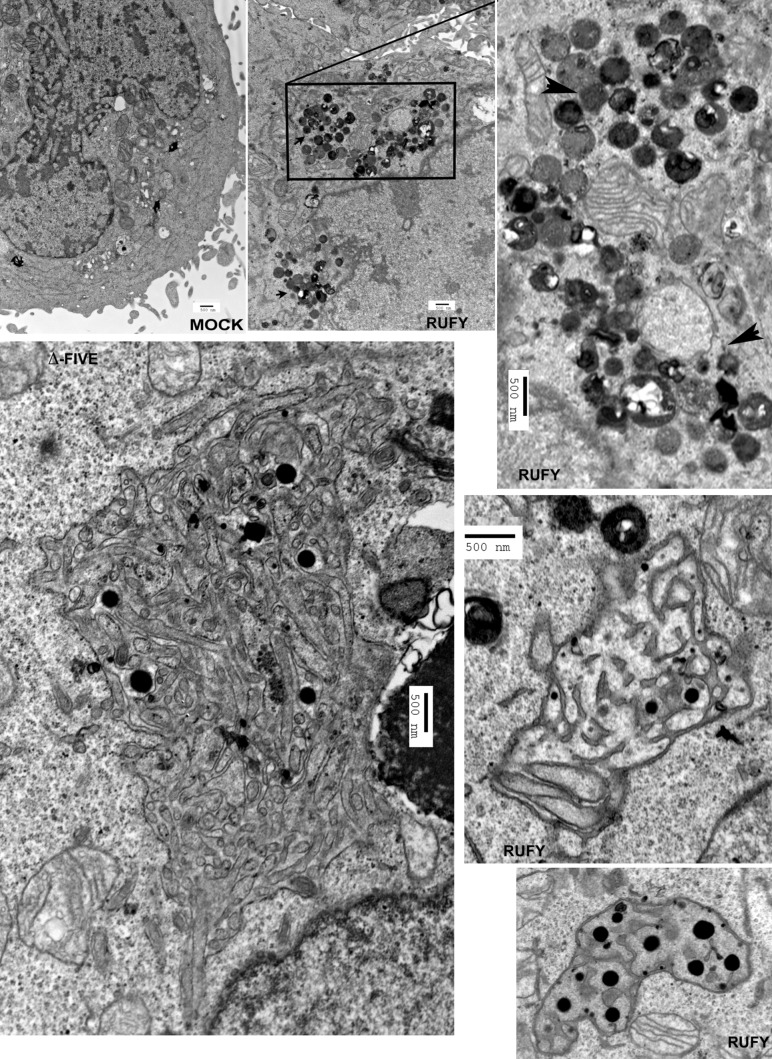
Figure 8.
Electron microscopy analysis of Rufy4-overexpressing HeLa cells. HeLa cells were transfected with human Rufy4 or ΔFYVE Rufy4 construct or with mock plasmid as control and were fixed with glutaraldehyde. Subcellular structures were analyzed by conventional electron microscopy. Boxed area is magnified on the right. Arrowheads indicate zones of lysosome clustering and tethering. Bars, 500 nm.

