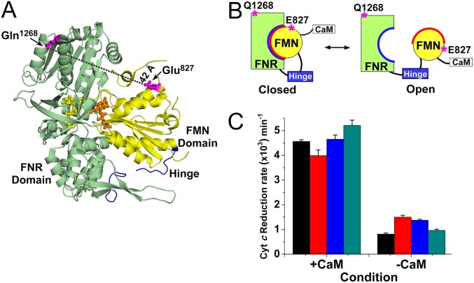
Fig. 1.
(A) The nNOSr ribbon structure (from PDB: 1TLL) showing bound FAD (yellow) in FNR domain (green), FMN (orange) in FMN domain (yellow), connecting hinge (blue), and the Cy3–Cy5 label positions (pink) and distance (42 Å, dashed line). (B) Cartoon of an equilibrium between the FMN-closed and FMN-open states, with Cy dye label positions indicated. (C) Cytochrome c reductase activity of nNOSr proteins in their CaM-bound and CaM-free states. Color scheme of bar graphs: Black, WT nNOSr unlabeled; Red, Cys-lite (CL) nNOSr unlabeled; Blue, E827C/Q1268C CL nNOSr unlabeled; and Dark cyan, E827C/Q1268C CL nNOSr labeled.

