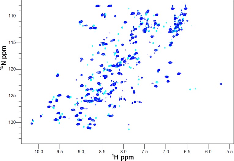
Fig. S10.
Electron transfer from Cu(I)-Sco2 to Apo-COX II*S-S followed by NMR. 1H-15N HSQC spectra of Apo-COX II*S-S (cyan) and upon addition of one equivalent of Cu(I)-Sco2 (blue). The disappearance of several cross peaks compared with the oxidized form of the protein is typical of spectra of COX II*2SH.

