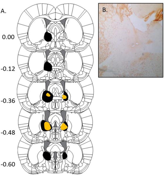
Figure 1.

BNST Lesions. A. Extent of the largest (black) and smallest (orange) BNST lesions accepted for analysis, ranging from +0.00 to −0.60 mm from bregma. From The Rat Brain in Stereotaxic Coordinates (6th ed.), Pages 33–38, by G. Paxinos & C. Watson, 2007, New York, NY: Academic Press. Copyright 2005 by Elsevier Academic Press. Adapted (or reprinted) with permission.B. Example of a BNST lesion in a NeuN stained brain slice.
