Figure 4.
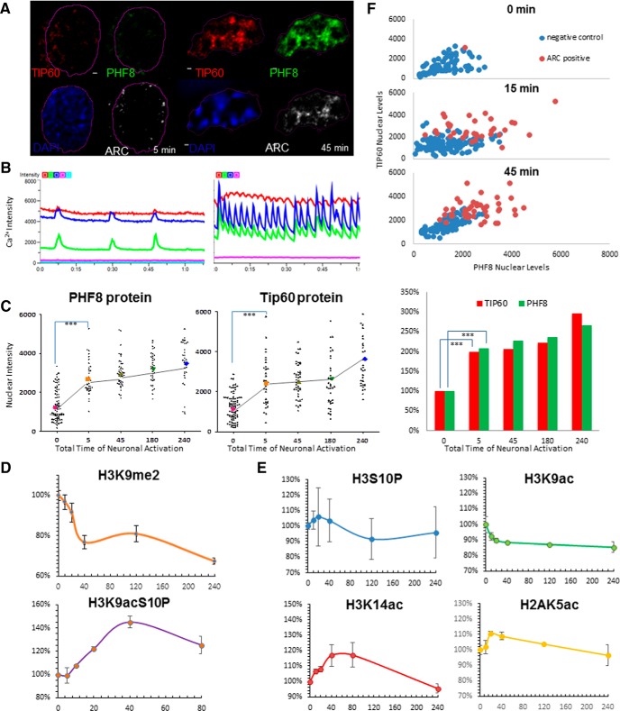
Neuronal activity reorganizes PHF8 and TIP60 in the nucleus and effectuate histone methylation and acetylation changes. A, A representative image of a pair of hippocampal neuronal nuclei during the first 5 min of 4AP+Bic+Fors treatment and then at 45 min, showing the activity-dependent increase of PHF8 and TIP60 protein in the nucleus. B, Neural network activity visualized by Ca2+ imaging (gCamp6 intensity over time), with each different-colored line representing individual neurons, before (left) and after (right) treatment with 4-AP+Bic. C, Dot plot of nuclear levels of PHF8 and TIP60 (unpaired t test; *** p ≤ 0.0001; one-way ANOVA: F = 33.23, R 2 = 0.3693). Each symbol represents the intensity of PHF8 or TIP60 staining from a single neuronal nucleus, lines correspond to the mean and SEM of all neurons imaged at the indicated time points. D, mRNA levels of PHF8 show a biphasic peak with time of chemLTP, whereas TIP60 shows an initial upregulation but a return to baseline within 45 min of sustained activity. E, Time-courses of chromatin modification of neurons imaged using high-content screening (n = 500 − 1000/site, 6 sites/well, 96-well; ImageXpress Micro, Molecular Devices) show an activity-dependent decrease in the overall per-nucleus intensity of H3K9me2 staining in neural networks treated with chemLTP, which coincides with a robust increase in H3K9acS10P. F, Graphs of nuclear PHF8 as a function of nuclear TIP60 levels at 0, 5, and 45 min of neural network activation in ARC-positive versus ARC-negative neurons, showing two identifiable distinct populations of neurons. The bar graph indicates levels of TIP60 (red) and PHF8 (green) as a function of time of synaptic activation (in minutes; y-axis). PHF8 and TIP60 are both highly induced within as early as 5 min of synaptic activation (p = 0.00001).

