
Figure 6.
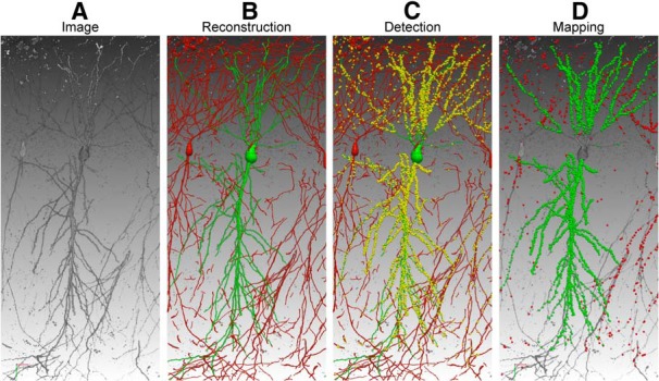
Mapping brain connectivity with neuTube 1.0. A, The original 3D confocal image contains postsynaptic neurons. B, The target neuron (green) was traced semi-automatically. The red branches belong to background neurons. C, mGRASP-labeled synapses (yellow) were detected automatically, with sizes enlarged for better visualization. D, The synapses were mapped to the target neuron (green) and background neurons (red).