Figure 1.

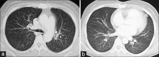
Contrast-enhanced CT axial images in lung window settings through upper (a) And lower lobes (b) Reveal randomly distributed miliary nodules in the both lungs

Contrast-enhanced CT axial images in lung window settings through upper (a) And lower lobes (b) Reveal randomly distributed miliary nodules in the both lungs