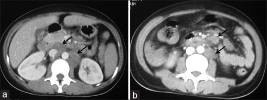
Figure 2.

Contrast-enhanced CT axial images at the level of renal hilum (a) And infra-renal levels. (b) Reveal discrete necrotic enlarged lymph nodes in aorto-caval, para-aortic locations and in the mesentery

Contrast-enhanced CT axial images at the level of renal hilum (a) And infra-renal levels. (b) Reveal discrete necrotic enlarged lymph nodes in aorto-caval, para-aortic locations and in the mesentery