Figure 1.

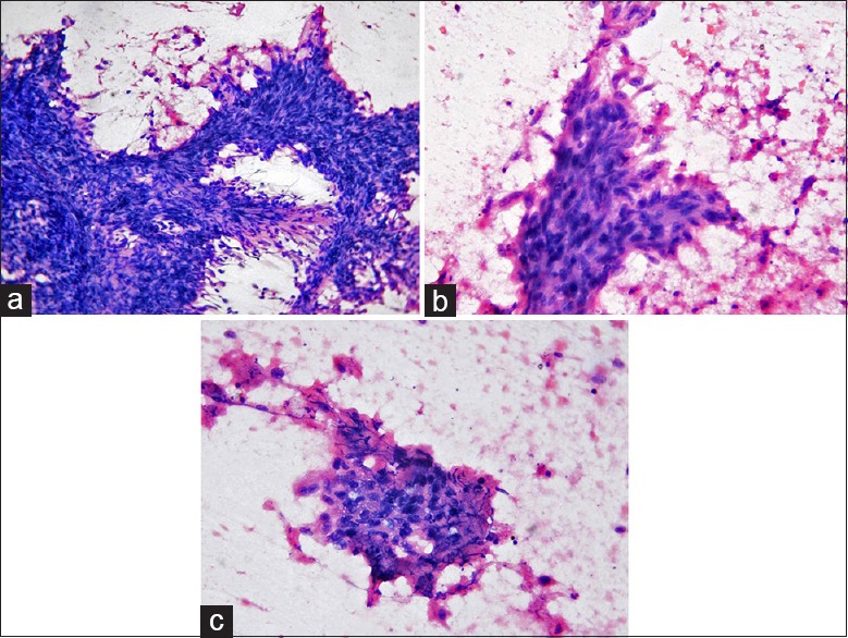
Aspiration cytology of the tumor: Photomicrographs show a malignant tumor composed predominantly of fragments of spindle-shaped cells (a) (HE, ×200) showing nuclear pleomorphism (b) (HE, ×400); few clusters of atypical epithelial cells are seen along with necrosis (c) (HE, ×400)
