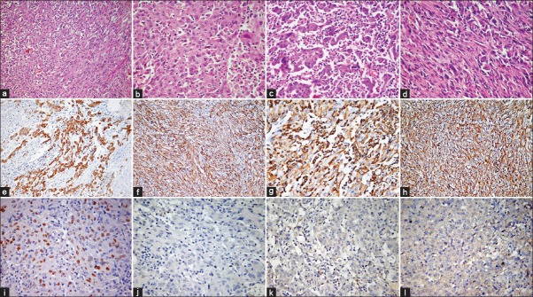
Figure 2.

Histopathological features: Photomicrographs showing a biphasic tumor with epithelial and spindle cells (a) (HE, ×200); epithelial component shows nuclear pleomorphism, frequent mitoses (b) (HE, ×400) and inflammatory infiltrate (c) (HE, ×400); sarcomatoid component shows fascicles of malignant spindle cells (d) (HE, ×400); immunohistochemistry for cytokeratin is positive in epithelial (e) and spindle cells (f) vimentin is positive in epithelial (g) and spindle cells (h) (IHC, ×200). TTF1 shows nuclear positivity (i) while p40 (j) EGFR E746-A750del (k) and L858R EGFR (l) are negative (IHC, ×400)
