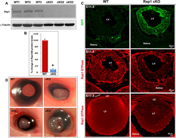
Fig. 2.

Ocular phenotype in the Rap1 cKO mouse. A & B. Intact lenses (membrane-enriched fraction) derived from three representative E17.5 Rap1 cKO mouse embryos showed a dramatic reduction (by ~ 90%; *P<0.05; n=4) of Rap1 (Rap1a/Rap1b-total) protein compared with WT controls based on immunoblot quantification. C. Reduction of Rap1 in lenses from E11.5 and E17.5 Rap1 cKO mouse embryos based on immunofluorescence analysis. The top panel shows the distribution profile of GFP (green fluorescent protein) only in the lens vesicle of Rap1 cKO mouse embryos (E11.5) confirming Cre expression relative to the WT control. The middle and lower panels show reduction of Rap1 in the E11.5 lens vesicle and E17.5 lens, respectively, derived from Rap1 cKO mice and relative to the WT controls based on immunofluorescence analysis. D. Conditional deletion of Rap1 (Rap1a/Rap1b) in the lens results in microphthalmic eyes with lens opacity in the embryonic and neonatal mice. The images shown are with eye lids removed from the E17.5 embryos. Bars in panel C represent image magnification. LV: Lens vesicle; LE: Lens epithelium; LF: Lens fibers.
