Fig. 4.


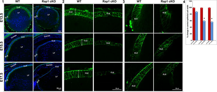
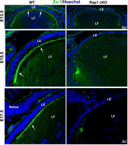
Disruption of E-cadherin-based AJs and ZO-1associated cell-cell interactions in Rap1 cKO mouse lenses. A. The Rap1 cKO mouse specimens derived from E13.5, 15.5 and 17.5 embryos reveal a progressive disruption in E-cadherin based AJs (green) in lens epithelium associated with altered cell shape and significantly reduced cell width compared to the respective WT controls (A1, A4). While lens epithelial cells (in both central (A2) and equatorial (A3) epithelium) in WT specimens exhibit a long columnar shape (double headed arrow; known to depend on maintenance of apical to basal polarity) and intense E-cadherin-positive AJs, the Rap1 cKO lens specimens exhibit a dramatic reduction in E-cadherin-based AJs both in central (A2) and equatorial (A3) epithelium, and disruption of apical to basal polarity with altered cell shape. A4 shows a significant decrease in lens central epithelial width in the Rap1 cKO specimens compared with WT, based on values (mean ± SEM) derived from 6 independent specimens. *P<0.05. LE: Lens epithelium, LF: Lens fibers, CLE: Central lens epithelium, ELE: Equatorial lens epithelium. B. Similar to AJs shown in Fig. 4A, ZO-1-based cell adhesive interactions were dramatically reduced in the lens of E13.5, E15.5 and E17.5 Rap1 cKO embryos. ZO-1 based cell-cell interactions (in green) were distributed discretely between the apical junctions of fiber cells and epithelium in the WT lenses (arrows). However, in the absence of Rap1, the ZO-1 based cell-cell interactions were dramatically reduced. The loss of cell adhesive interactions was associated with reduction in number and size of nuclei in the lens epithelium of Rap1 cKO specimens relative to WT controls, based on Hoechst staining (blue). Bars represent magnification.
