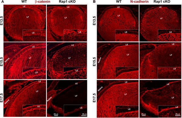
Fig. 5.

Rap1 deficiency suppresses β-catenin-based cell-cell junctions and increases N-cadherin levels in mouse lens epithelium. A. β-catenin, a well-characterized component of AJs localizes to the cell-cell junctions of lens epithelium and fiber cells in WT specimens based on immunofluorescence analysis (red) of paraffin embedded sagittal sections. In Rap1 cKO E13.5, E15.5 and E17.5 specimens, there is a progressive and dramatic reduction of β-catenin staining in both lens epithelium and fibers compared to WT specimens. The insets show magnified areas of lens epithelium. B. Unlike β-catenin, N-cadherin distribution (red staining) is relatively intense in fiber cells compared to the epithelium of WT lenses of mouse embryos. However, in the Rap1 cKO mouse lens specimens (E13.5, 15.5 and 17.5), there was a progressive and marked increase in N-cadherin staining in the epithelium with a dramatic and concomitant reduction in the fiber cells. The insets show the magnified area of lens epithelium. Bars represent image magnification. LE: Lens epithelium, LF: Lens fibers.
