Fig. 7.


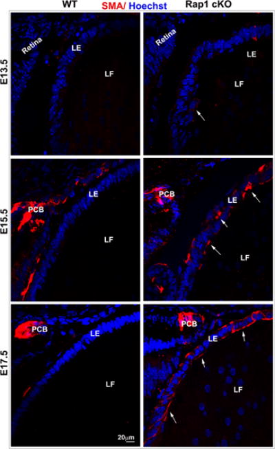
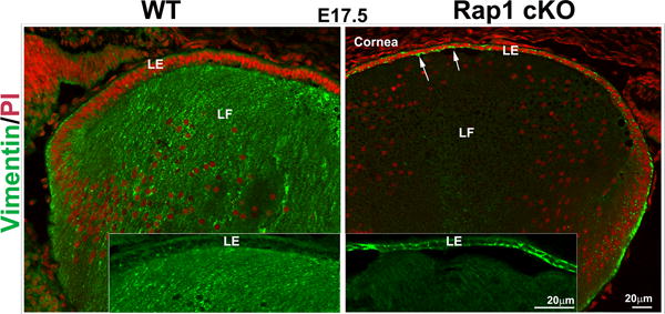
Rap1 deficiency induces EMT in mouse lens. A. To determine the effects of Rap1 deficiency on lens epithelial plasticity, E13.5, 15.5 and 17.5 ocular specimens derived from Rap1 cKO mouse embryos were evaluated for changes in αSMA by immunofluorescence analysis using an anti-αSMA monoclonal antibody conjugated with Cy3™. While the respective WT specimens show absence of αSMA in lens epithelium, the Rap1 cKO mouse lens specimens exhibit progressively increased levels of αSMA in the epithelium of E13.5, E15.5 and E17.5 specimens (indicated with arrows; red staining). Both WT and Rap1 cKO mouse specimens exhibit αSMA staining in the presumptive ciliary body (PCB) and iris. B. In addition to αSMA, changes in vimentin were examined in the Rap1 cKO mouse ocular specimens by immunofluorescence analysis. In E17.5 WT specimens, fibers stain intensely for vimentin (green) with very little positive staining in the lens epithelium (see inserts). Rap1 cKO mouse lens specimens in contrast, exhibit a marked increase in vimentin staining in the lens epithelium (arrows), with a concomitant decrease in the fiber cells. Insets show magnified areas of lens epithelium and fibers. Red staining shows propidium iodide (PI)-based nuclei distribution. LE; Lens epithelium, LF: Lens fibers. Bars in A and B represent image magnification.
