Fig. 9.


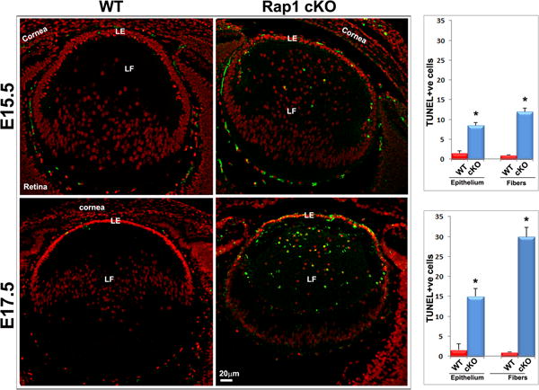
Rap1 deficiency impairs lens epithelial proliferation and survival. A. To determine the effects of Rap1 deficiency on lens epithelial proliferation and cell cycle progression, in vivo BrdU labeling was performed in conjunction with immunofluorescence analysis using anti-BrdU antibody as described in Methods section. Counting of BrdU positive cells showed a significant decrease (>50%) in the lens central epithelium of Rap1 cKO embryos relative to their WT controls. In contrast to the lens central epithelium, no BrdU-positive cells are noted in the transitional zone of the WT epithelium, where cells exit from the cell cycle and start differentiating into secondary fiber cells (a region just below the line drawn and indicated with arrows). Rap1 cKO mouse lens specimens on the other hand, exhibited a significant increase in the number of BrdU positive cells in the transitional zone epithelium (arrows) indicating defective cell cycle exit in the deficiency of Rap1. B. To test the effects of Rap1 deficiency on lens epithelial and fiber cell survival, we examined for changes in apoptotic cells by TUNEL positive staining of ocular specimens in E15.5 and E17.5 mouse embryos. The Rap1 cKO mouse specimens showed a progressively and significantly increase of apoptotic cells (TUNEL positive cells- in green/yellow) in the lens epithelium and fiber mass compared with WT controls. Values (mean ± SEM) were based on n=6. *P<0.05. Bars show image magnification. LE: Lens epithelium, LF: Lens fibers.
