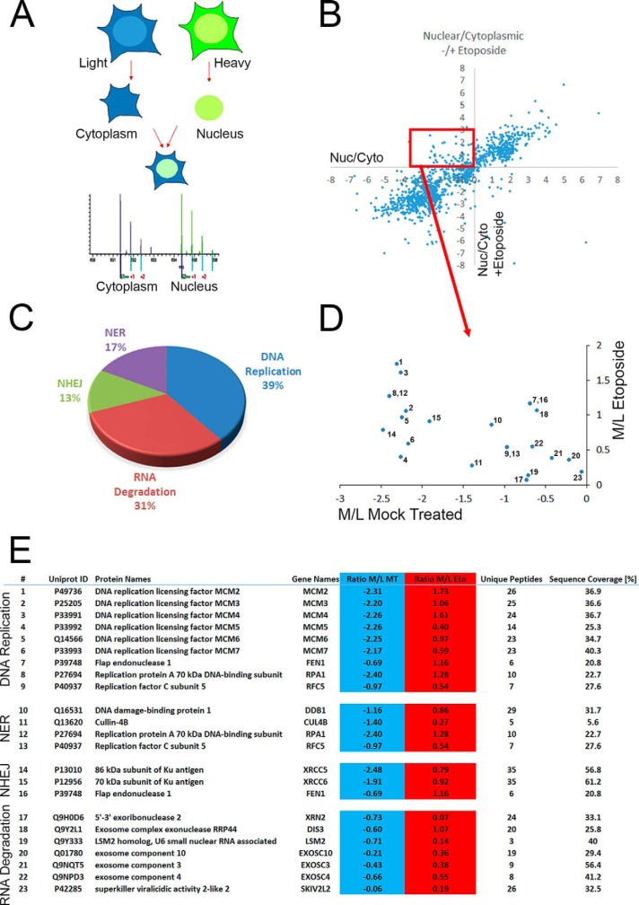
Fig. 1.
Protein subcellular relocalisation following DNA damage. A, Schematic diagram of spatial proteomics to quantify cytoplasmic and nuclear localization. Proteins from SILAC-labeled cells are biochemically fractionated into cytoplasmic and nuclear fraction which are then recombined prior to identification and quantification by mass spectrometry. B, Nuclear/Cytoplasmic ratios of proteins from mock treated cells (x axis) compared with proteins treated with 50 μm Etoposide for 1 h (y axis). C, KEGG pathway enrichment analysis on the 93 proteins showing a cytoplasmic to nuclear relocalisation following DNA damage. D, Numeration of the 23 proteins identified in the KEGG pathway enrichment according to the table (E) showing the list of proteins classified in each KEGG pathways.

