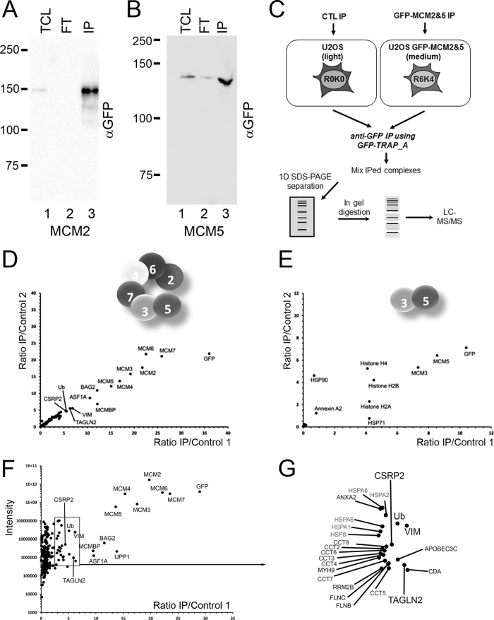
Fig. 3.
AP-MS of GFP-MCM2 and GFP-MCM5 to identify interacting proteins. A, GFP tagged MCM2 and B, MCM5 proteins were immunoprecipitated using GFP-TRAP agarose beads, ensuring a near depletion of all the tagged MCM protein from the cell lysate. C, SILAC-labeled cells were used for comparison of GFP-based immunoprecipitates from uninduced cells (light) with doxycycline-induced cells (medium). Immunoprecipitates were combined, separated by SDS-PAGE, each gel lane cut into eight slices prior to in-gel digestion with trypsin. The extracted peptides were analyzed by LC-MS/MS. The M/L ratio of two independent experiments for GFP-MCM2 (D) and GFP-MCM5 (E) were plotted and proteins with ratio above the level of the contaminants were identified. F, The average M/L ratios of the two GFP-MCM2 AP-MS experiments were plotted versus the total intensities. G, A zoom over the boxed region in F, for identification of proteins with ratios above the contaminants.

