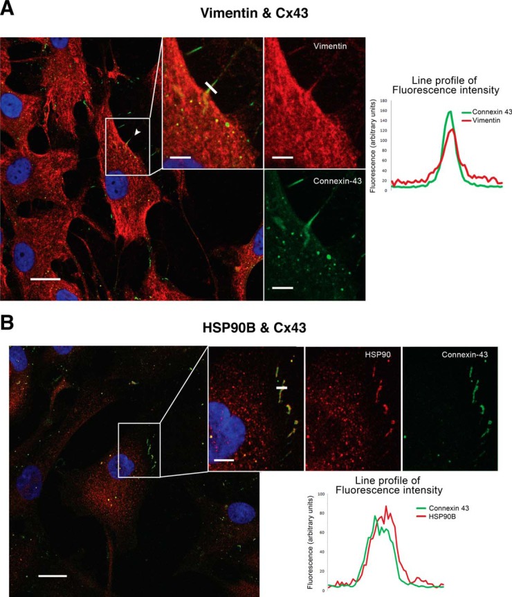
Fig. 3.
Colocalization of Cx43 with vimentin and HSP90β in articular chondrocytes. Immunofluorescence confocal microscopy was used to study colocalization of two antigens in one cell. A, Images represent the double staining and DAPI in chondrocytes in monolayer culture that were obtained from healthy human donors. Cx43 is detected in green, and vimentin is in red. B, Colocalization between HSP90β and Cx43 was also studied in human chondrocytes in monolayer culture. Cx43 is shown in green, and HSP90β is in red color. Right panels; colocalization graphs show the fluorescence intensity profile from a line crossing through the cell protrusions, the coincidence of fluorescence intensity peaks, for both red and green, marks the colocalization of both proteins.

