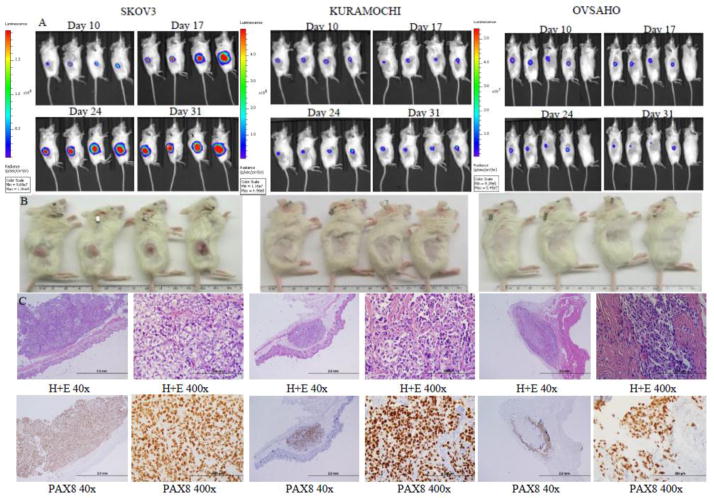
Figure 3.
Xenograft growth of cell lines after subcutaneous implantation. A) Tumor growth by bioluminescent imaging. B) Photographs of tumors after euthanasia. C) Light microscopy images of tumors at 40x and 400x magnification showing histology by hematoxylin and eosin staining and identification of tumor cells by PAX8 immunohistochemistry.

