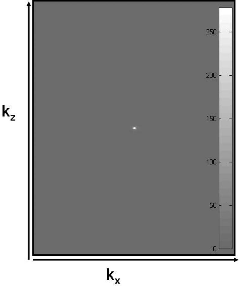
Figure 1.
k-space data (absolute value) of a 321 slice acquisition of a human brain using a 3D FLAIR sequence. (Data along ky was MIPed to project the data in two dimensions.) kx has 256 steps while kz has 321 overcontiguous steps. Embedded gray scale bar shows that the window was adjusted to accomodate the entire range of data values (≥ 0).

