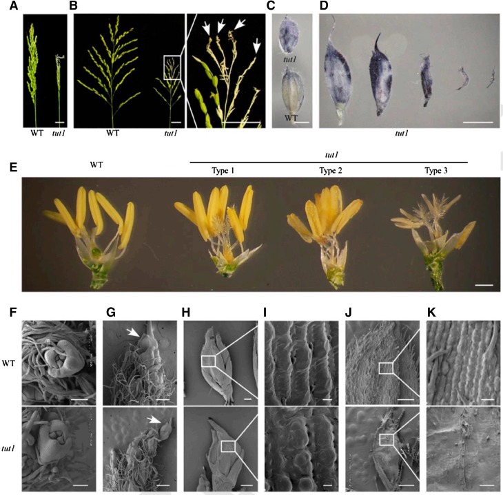
Figure 2.
Morphology of panicle and spikelet in tut1. A, Panicle rachis of wild-type (WT; left) and tut1 (right) plants at the grain-filling stage. Bar = 2 cm. B, The expanding panicle branch of wild-type (left) and tut1 (right) plants at the grain-filling stage. The part of the tut1 panicle shown in the white box was enlarged. The arrows show the degenerated branches and spikelets in the tut1. Bars = 2 cm. C, ROS analysis of the wild type (bottom) and tut1 (top) by NBT staining. Bar = 2 mm. D, ROS analysis of spikelets showing different degrees of degeneration in tut1 at the heading stage by NBT staining. Bar = 2 mm. E, Comparison of wild-type (left one) and tut1 spikelets (right three) after removing the lemma and palea at the heading stage. Right three spikelets, named Type 1, Type 2 and Type 3, show varying degrees of developmental defects in tut1. Bar = 1 mm. F to K, Scanning electron microscopy (SEM) observation of spikelet at the tip of the panicle at stage In7 (F), stage In8 (arrows indicate the apical spikelets in the wild type and the tut1 [G]), stages In8 and In9 (H; the white square frame region shown in H is enlarged in I), and stage In9 (J; the white square frame region shown in J is enlarged in K). Bars = 100 μm (F and K), 400 μm (G), 200 μm (H), 10 μm (I), and 500 μm (J).

