Figure 3.
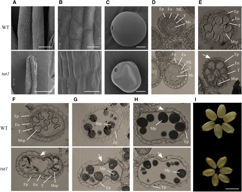
SEM and semithin section analysis of wild-type and tut1 anther morphology. A to C, SEM observation of mature anthers of the wild type (WT) and tut1 (A), enlarged view of the anther surface of the wild type and tut1 (B), and mature pollen grain of the wild type and the tut1 (C). Bars = 200 μm (A), 20 μm (B), and 10 μm (C). D to H, The cross sections were stained with toluidine blue. Ep, Epidermis; En, endothecium; ML, middle layer; Ms, microsporocyte; Msp, microspore; Mp, mature pollen; T, tapetum. Cross section of single locule at the microspore mother cell stage; Ep, En, ML, T, and Ms are indicated (D); vacuolated pollen stage; Ep, En, T, and Msp are indicated (E); pollen first mitosis stage; Ep, En, T, and Msp are indicated (F); pollen second mitosis stage; Ep and Mp are indicated (G); and mature pollen stage; Ep and Mp are indicated (H). The arrows show the degenerated epidermal cell. I, Phenotype of mature seeds without glumes of the wild type (top) and tut1 (bottom). Bar = 5 mm.

