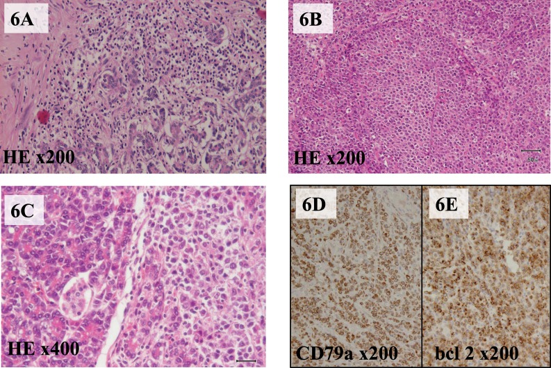
Fig. 6.
(A) Microscopic examination following hematoxylin-eosin staining, which showed acute pancreatitis with lymphocyte-infiltrating parenchyma of the pancreas tail. (B) Hematoxylin-eosin staining, which demonstrated follicular-type lymphoma cells. (C) Lymphoma cells involving the parenchyma of the exocrine pancreas and Langerhans islets. Both CD79a immunostaining (D) and bcl-2 immunostaining (E) are positive.

