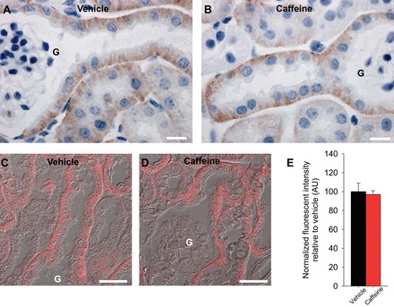
Fig. 8.

In the mouse kidney NBCe1 is localized to the basolateral membrane of S1 and early S2 segments of the proximal tubule. Representative images of diaminobenzidine-stained kidneys shows basolateral NBCe1 abundance in the S1 and early S2 segment of the proximal tubule (A), and caffeine application did not clearly change this staining pattern (B). C and D: representative differential interference contrast overlayed quantitative laser scanning microscopy images of NBCe1 in S1 proximal tubule cells. Mean fluorescent signal intensity of NBCe1 labeling was not significantly different between vehicle- and caffeine-treated mice. G, glomerulus; n = 6/condition. Scale bar = 10 μm (A and B) and 25 μm (C and D).
