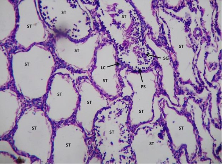
Figure 7.
Light microphotograph (H&E stained) section of sodium arsenite treated testicular section showing severe degeneration. Seminiferous tubules with no spermatogenetic stages or if are present, only 5% denotes the normal functioning. The leydig cells are in highly degenerative condition as hemorrhage in them is observed x400.

