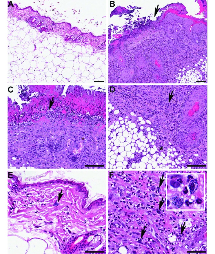
Figure 1.
Representative histologic appearance of UD. (A) Normal skin from an unaffected mouse. Note the thin epidermis and dermis above abundant subcutaneous fat. (B) A focus of ulcerative dermatitis in an affected animal, imaged at the same magnification as in A. The arrow denotes the junction between intact, markedly hyperplastic and hyperkeratotic epidermis (on the right) and a locally extensive area of ulceration (on the left). The ulcerated area is subtended by inflammation extending into the deep dermis and subcutis. (C) Higher magnification image of the area of ulceration, showing extensive serocellular crusting with numerous degranulated neutrophils (arrow) accompanying epidermal necrosis. (D) High magnification of the deeper portions of the ulcerated area, showing mixed mononuclear and granulocytic inflammation (arrow) in the dermis and extending into the subcutis (asterisk). (E) High magnification of normal skin, showing lack of inflammation in the dermis, with presence of occasional mast cells (arrow). (F) High magnification of the dermis in an animal with ulcerative dermatitis showing that the inflammatory infiltrate contains numerous mast cells (arrows, and inset). Hematoxylin and eosin stain; bar, 100 μm (A through D); 50 μm (E and F).

