
Figure 3.
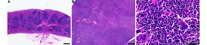
Representative histologic appearance of spleens from mice (A) without and (B) with UD. (A) Histologic appearance of a normal spleen in a mouse without dermatitis. (B and C) Histologic appearance of enlarged spleen in a mouse with ulcerative dermatitis. The architecture of the normal spleen is altered, in that (B) there are no well-defined lymphoid areas, and (C) the red pulp sinuses are markedly expanded by extramedullary hematopoietic elements. Hematoxylin and eosin stain; bar, 100 µm (A and B), 50 µm (C).