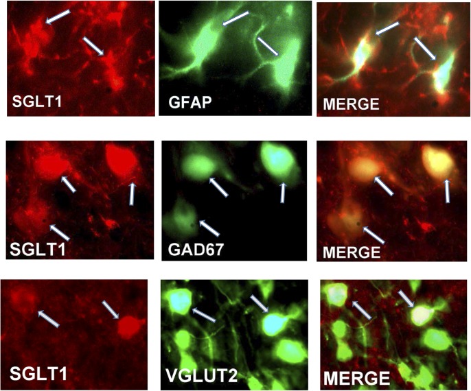
Figure 5.
Top panels: Immunohistochemical staining for SGLT1 (red), the astrocytic marker GFAP (green), and the merged image showing colocalization of SGLT1 with GFAP (arrows). Middle and bottom panels: Immunohistochemical staining for SGLT1 (red), the neuronal markers GAD67 and VGLUT2 (green), and the merged image showing colocalization of SGLT1 with GAD67 and VGLUT2 (arrows).

