
Fig. 1.
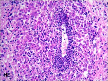
Histopathologic examination evidenced an angiocentric inflammatory lesion in the cerebral white matter composed of macrophages and mixed with lymphocytes and plasma cells (hematoxylin and eosin, ×400)
Official websites use .gov
A
.gov website belongs to an official
government organization in the United States.
Secure .gov websites use HTTPS
A lock (
) or https:// means you've safely
connected to the .gov website. Share sensitive
information only on official, secure websites.
Histopathologic examination evidenced an angiocentric inflammatory lesion in the cerebral white matter composed of macrophages and mixed with lymphocytes and plasma cells (hematoxylin and eosin, ×400)